Spotlight on Skin Cancer Procedures: A Precise Approach to Skin Cancer Treatment

Skin cancer is a serious health concern affecting millions of people worldwide. In Australia, where sun exposure is prevalent, it’s particularly significant. Understanding skin cancer, its types, and symptoms is crucial for early detection and effective treatment. With higher sun exposure in Australia, it is essential that one should be able to distinguish difference between normal skin spot and a cancerous one. This article aims to shed light on different skin cancers and their traits.
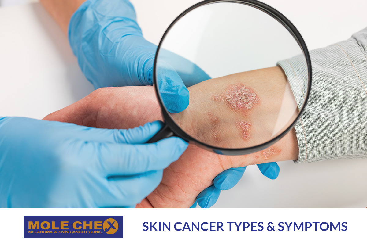
The Three Main Types of Skin Cancers :
Basal Cell Carcinoma (BCC)
This is the most common type, accounting for major number of skin cancers. BCC starts in the basal cells of the skin and usually grows slowly over months or years. While it rarely spreads to other parts of the body, untreated BCCs can invade nerves and surrounding tissue, making treatment more challenging.
Signs of Basal Cell Carcinoma (BCC):
- Development on Sun-Exposed Areas: BCC often appears on areas of the body that receive significant sun exposure, such as the head, face, neck, shoulders, lower arms, and legs. However, it can occur anywhere on the body.
- Pearl-Colored Lump or Scaly Area: BCC typically presents as a raised, pearl-colored lump or a slightly scaly area on the skin. This area may appear shiny and pale or bright pink. In some cases, BCCs can appear darker.
- Skin Breakdown (Ulceration): If left untreated, BCCs can grow deeper into the skin. This can lead to the skin breaking down or ulcerating, causing the lesion to bleed and become inflamed. Sometimes, BCCs seem to heal but then become inflamed again.
- Slow Growth: BCC usually grows slowly over months or years. While it’s a less aggressive form of skin cancer, it can become more challenging to treat if it invades nerves and damages nearby tissues.
- Multiplicity: Having one BCC increases the risk of developing another. It’s not uncommon for individuals to have multiple BCCs simultaneously on different parts of their bodies.
Squamous Cell Carcinoma (SCC)
SCC is the second most common type, comprising approximately 33% of skin cancers. Unlike BCC, SCC can grow quickly over several weeks or months. Some SCCs remain on the skin’s top layer, while invasive SCC can spread to other parts of the body.
Signs of Squamous Cell Carcinoma (SCC):
- Quick Growth: SCCs can grow rapidly over several weeks or months, which distinguishes them from BCCs that tend to grow more slowly.
- Top Layer or Invasive: Some SCCs are confined to the top layer of the skin. These are known as “SCC in situ,” “intra-epidermal carcinoma,” or “Bowen’s disease.” However, if SCC invades through the basement membrane, it’s classified as “invasive SCC.” Invasive SCC has the potential to spread to other parts of the body, a condition known as “metastatic SCC.”
- Location Matters: SCC on certain parts of the body, like the lips and ears, is more likely to metastasize or spread to other areas.
- Change in Appearance: SCCs may initially appear as scaly, rough, or slightly raised areas on the skin. Over time, they can change in appearance and become more noticeable.
- Potential for Spreading: SCCs have a higher potential to spread than BCCs. Early detection and treatment are crucial to prevent metastasis and ensure a better prognosis.
Melanoma
Although less common than BCC and SCC, melanoma is the deadliest form of skin cancer. It originates in melanocytes, the pigment-producing cells. Melanoma can spread rapidly to other organs if not detected early.
Signs of melanoma, can be identified using the ABCDEs of melanoma detection. These criteria help individuals and healthcare professionals assess whether a mole or skin lesion may be melanoma. Here are the signs to look for:
- Asymmetry: One half of the mole or lesion does not match the other half. In other words, if you were to draw a line through the middle, the two halves would not be identical in shape.
- Border Irregularity: The edges of the mole are uneven, ragged, or notched. Melanomas often have irregular, poorly defined borders compared to benign moles, which typically have smooth, even borders.
- Color Variation: Melanomas can have multiple colors within the same lesion. This may include shades of brown, black, tan, red, blue, or white. Benign moles are usually a single, uniform color.
- Diameter: Melanomas are typically larger in diameter than common moles. While melanomas can be smaller, they are often larger than 6 millimetres (about the size of a pencil eraser) when diagnosed.
- Evolution or Change: Any change in size, shape, color, or symptoms (such as itching, bleeding, or tenderness) of a mole or skin lesion should be taken seriously. Be particularly vigilant if a mole starts to evolve or exhibit any of the above changes.
It’s essential to remember the “E” in the ABCDEs, which stands for “Evolution” or “Change.” If you notice any changes in a mole or skin lesion, consult a dermatologist promptly. Early detection and treatment of melanoma can greatly improve outcomes.
Additionally, it’s important to recognize the “Ugly Duckling” sign, which refers to a mole or lesion that looks significantly different from the person’s other moles. If you have a mole that stands out due to its appearance, consult a healthcare professional for evaluation.
Regular self-examinations and annual skin checks by a dermatologist are crucial for detecting melanoma and other skin cancers early, when they are most treatable.
It’s essential to consult a dermatologist if you notice any unusual changes in your skin, such as new growths, changes in the appearance of existing moles or spots, or any persistent skin issues. Early diagnosis and intervention can significantly improve outcomes for both BCC and SCC.
Other Skin Spots and Conditions
- Sunspots (Actinic or Solar Keratoses): These flat, scaly spots are often a sign of excessive sun exposure and can sometimes develop into SCC.
- Moles (Naevi): Moles are common and usually harmless. However, individuals with numerous moles have a higher risk of melanoma.
- Dysplastic Naevi: Irregular moles are linked to an increased risk of melanoma.
- Age Spots (Seborrhoeic Keratoses): These common growths are usually harmless but may resemble skin cancer.
Sunspots:
Sunspots, also known as actinic keratoses or solar keratoses, are common skin growths that can develop due to prolonged exposure to ultraviolet (UV) radiation from the sun. These spots are considered precancerous because they can sometimes progress to skin cancer, particularly squamous cell carcinoma (SCC), if left untreated. Here’s a more detailed explanation of sunspots and their signs:
Sunspots are rough, scaly patches or lesions that typically develop on areas of the skin that have been exposed to the sun, such as the face, neck, hands, forearms, and legs. They are a result of UV damage to the skin’s DNA, which causes abnormal cell growth. Sunspots can vary in size, color, and texture. They are often small, measuring a few millimetres in diameter, but they can become larger if left untreated.
Signs of Sunspots:
- Appearance: Sunspots often appear as flat, scaly, or crusty spots on the skin. They can range in color from the same color as your skin to red. Some may appear reddish or pinkish in the early stages.
- Texture: They typically have a rough or gritty texture. You may feel the roughness when you touch them.
- Ease of Removal: Sunspots can sometimes be easily scratched off, but they tend to return within a few days to weeks.
- Persistent Changes: If left untreated, sunspots can persist and may develop into skin cancer, particularly SCC.
It’s important to note that while sunspots are considered precancerous, not all of them will progress to skin cancer. However, because some can become cancerous over time, it’s essential to have any suspicious skin growths or changes evaluated by a dermatologist. Early detection and treatment of sunspots can help prevent them from developing into skin cancer.
Moles (naevi)
Skin Moles, also referred to as naevi (singular: nevus), are common skin growths that form when melanocytes, the pigment-producing cells in the skin, grow in clusters instead of spreading evenly. Moles can appear anywhere on the body and vary in size, shape, color, and texture. They are usually harmless, but in some cases, they can develop into melanoma, a type of skin cancer. Here’s an in-depth explanation of moles and their signs:
Characteristics of Moles:
- Color: Moles can be brown, black, tan, red, pink, or flesh-colored.
- Shape: They can be round or oval.
- Texture: Most moles are smooth and flat. Some may be slightly raised.
- Size: Moles can range in size from tiny dots to larger than a pencil eraser.
- Borders: Moles often have well-defined, clear borders that distinguish them from the surrounding skin.
- Location: They can appear anywhere on the body, including areas not exposed to the sun.
Dysplastic Nevi (Atypical Moles):
Dysplastic nevi, often referred to as atypical moles, are a unique category of moles that differ from typical moles in appearance and potential risk. These moles are associated with a higher risk of developing melanoma, a serious form of skin cancer.
Signs of Dysplastic Nevi (Atypical Moles)
- Irregular Shape: Dysplastic nevi typically have an irregular or asymmetrical shape. Instead of being perfectly round or oval, they may have uneven borders with notches, scalloped edges, or an irregular outline.
- Uneven Color: These moles often display uneven coloring. They can contain various shades of brown, pink, red, and tan, resulting in a mottled appearance.
- Size: Dysplastic nevi can be larger than ordinary moles, often exceeding the size of a pencil eraser (6 millimeters or more).
Age Spots (Seborrhoeic Keratoses):
Age spots, also known as seborrhoeic keratoses, are benign skin growths commonly found in individuals as they age. These growths can appear almost anywhere on the body except the palms of the hands and the soles of the feet. While they might resemble certain types of skin cancer or even sunspots, age spots are typically harmless. One important distinguishing feature of age spots is that they can be easily scratched, potentially leading to bleeding
Signs of age spots (seborrhoeic keratoses) can include:
- Raised Warty Texture: Age spots often have a raised, rough, or warty texture, which can make them feel different from the surrounding skin.
- Brown to Dark Brown Color: These growths typically appear in shades of brown, ranging from light to very dark brown.
- Nonuniform Surface: Age spots may have an uneven surface and can be slightly scaly.
- Variety of Sizes: They can vary in size, from small spots to larger, more noticeable growths.
- Round or Oval Shape: Most age spots are round or oval in shape, but they can sometimes be irregular.
- Itching: While usually not painful, age spots can occasionally become itchy or irritated.
- Bleeding: Scratching or rubbing an age spot might cause it to bleed, but this is generally not a serious concern.
Causes and Risk Factors
Skin cancer is primarily caused by exposure to UV radiation, which can come from the sun or artificial sources like tanning beds. High levels of UV radiation, prevalent in most parts of Australia, increase the risk of skin cancer. Other risk factors include fair skin, frequent sunburns, previous skin cancer diagnoses, family history, and certain skin conditions.
Over 95% of skin cancers are caused by UV radiation, primarily from the sun but also from artificial sources like tanning beds. Australia has high UV radiation levels year-round. Prolonged exposure can lead to sunburn, premature aging, and skin cell damage, increasing the risk of skin cancer. Risk factors include fair skin, red or light-colored hair and eyes, UV exposure, tanning bed use, outdoor work, weakened immune system, many moles or irregular moles, family history of skin cancer, and certain skin conditions. Darker skin offers some UV protection but doesn’t eliminate the risk.
Various factors can heighten the risk of developing skin cancer. While skin cancer can affect individuals of all ages, it becomes more prevalent with advancing age. Risk factors include having:
- Fair or freckled skin, especially if it is prone to easy burning and does not tan easily.
- Red or light-colored hair and pale eyes (such as blue or green).
- Prolonged and unprotected exposure to UV radiation, particularly in the form of short, intense periods of sun exposure and sunburn, often occurring during weekends and holidays.
- A history of active tanning or the use of solariums.
- Occupations or activities that involve prolonged outdoor exposure or contact with substances like arsenic.
- A weakened immune system, which can result from conditions like leukemia or lymphoma, or the use of immunosuppressive medications (for conditions such as rheumatoid arthritis, other autoimmune disorders, or organ transplants).
- Numerous moles or moles that exhibit irregular shapes and uneven colors.
- A prior history of skin cancer or a family history of the disease.
- Specific skin conditions, such as sunspots.
It’s worth noting that individuals with olive or very dark skin possess some natural protection against UV radiation due to their skin’s increased melanin production compared to fair skin. However, even individuals with darker skin tones can still develop skin cancer and should take precautions to protect their skin.
Conclusion : Skin Cancer Statistics in Australia
Australia has one of the world’s highest rates of skin cancer. Approximately two in three Australians will be diagnosed with some form of skin cancer before the age of 70. Non-melanoma skin cancer is the most common type in the country, with over one million treatments provided each year.
Knowing the types and symptoms of skin cancer is the first step in its prevention and treatment. Early detection can save lives. Stay informed and protect your skin from harmful UV radiation. Regular skin checks with a dermatologist can help identify potential issues before they become serious. Don’t take chances with your skin’s health. Remember, regular skin checks are a proactive step toward protecting your skin health. Schedule a skin checks today and ensure your skin’s well-being for the future.
Interested in learning more ? Visit skin cancer website : cancer.org.au
Read other article :


