
What Actually Happens If a Doctor Finds an “Atypical” Mole?
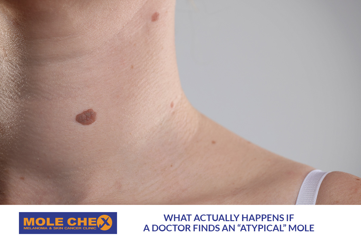
Finding an unusual mole during a skin check can create an immediate worry: “Does this mean skin cancer?” Fortunately, an atypical mole does not automatically mean melanoma. The term often describes a mole with features that differ from common moles. A doctor may examine it more closely, photograph it, monitor it or recommend a biopsy. The next step depends on how the mole looks, your personal risk and whether it has changed.
What Does “Atypical Mole” Actually Mean?
An atypical mole, also called an atypical naevus, may look different from an ordinary mole.
It might have an irregular shape, uneven border or several shades of colour. It may also appear larger or simply look different from your other spots.
Importantly, “atypical” describes appearance. It is not the same as a melanoma diagnosis. Many atypical moles prove to be benign. However, some can resemble melanoma closely enough that doctors need further assessment.
The term “dysplastic naevus” is also sometimes used. Strictly speaking, doctors usually make that diagnosis after examining tissue under a microscope.
What Happens During an Atypical Mole Check in Brisbane?
The first step usually involves looking carefully at the mole and surrounding skin.
A doctor may ask several questions. When did you first notice it? Has the size, shape or colour changed? Has it started bleeding, itching or crusting?
Your personal and family history can also matter. The doctor will often use a dermatoscope. This handheld device magnifies structures beneath the skin surface that are difficult to see with the naked eye.
Dermoscopy can help trained clinicians distinguish many harmless lesions from spots requiring closer investigation. Cancer Council Australia recommends dermoscopy for clinicians examining lesions for skin cancer detection. At Molechex, our Brisbane skin checks may include dermoscopic examination of suspicious spots. Our doctors have undertaken additional training in skin cancer medicine.
Does an Atypical Mole Always Need Removal?
No.
Some people expect every unusual mole to be removed immediately. That approach is not always necessary.
The doctor considers the mole’s appearance, dermoscopic features and history. They may also compare it with the rest of your moles.
People often develop their own general pattern of moles. A spot that looks very different from that pattern can attract greater attention. Doctors sometimes call this the “ugly duckling” concept.
When a lesion has no strongly suspicious features, a doctor may decide that monitoring is appropriate. Digital photographs or measurements can help document what the mole looks like today. Later comparisons may reveal subtle changes.
Australian guidance recognises sequential digital dermoscopic imaging as useful for selected lesions and patients.
Why Might a Doctor Recommend a Biopsy?
Sometimes appearance alone cannot provide enough certainty.
A doctor may recommend biopsy when a lesion has features that raise concern about melanoma or another skin cancer. A biopsy allows a pathologist to examine the cells under a microscope.
When melanoma is suspected, complete excision biopsy is generally preferred where practical. The doctor removes the lesion with a small surrounding margin under local anaesthetic. The tissue then goes to a pathology laboratory.
Other biopsy approaches may suit particular situations. The doctor chooses the method according to the lesion’s location, size and clinical features.
A biopsy recommendation does not mean cancer has already been diagnosed. It means the doctor needs more information to identify the lesion accurately.
What Happens on the Day of an Excision Biopsy?
For many patients, an excision biopsy is a relatively straightforward procedure.
The doctor first cleans the area and injects local anaesthetic. This numbs the skin around the mole.
The doctor then removes the lesion and usually closes the area with stitches. A dressing protects the wound while it begins healing. You will receive instructions about wound care, physical activity and when stitches need removal.
The removed tissue goes to pathology. A pathologist examines it microscopically and prepares a report.
That report provides much more information than visual examination alone.
What Can the Pathology Result Show?
There are several possible outcomes.
The mole may simply be benign. In that situation, no further treatment may be required.
The pathology report may describe a dysplastic or atypical naevus. Management then depends on the degree of atypia, whether the lesion was completely removed and the clinical circumstances.
Importantly, dysplastic naevi are not simply “melanomas waiting to happen”. Routine removal of every atypical mole does not prevent melanoma.
If pathology confirms melanoma, the next steps depend on features within the report. Your doctor will explain the diagnosis and recommended treatment. Further surgery may be needed to remove an appropriate margin of surrounding skin.
Some melanomas need additional investigation or specialist involvement. The exact pathway depends on the individual diagnosis.
What If the Doctor Decides to Monitor the Mole?
Monitoring does not mean ignoring the spot.
Instead, it means creating a comparison point and watching for meaningful changes. A doctor may record the lesion’s size or take a dermoscopic photograph. A later examination can then compare the same spot.
This approach can be particularly useful for people who have many moles. It can help identify changes without removing every unusual-looking but stable lesion.
However, you should not wait for the next planned review if the mole changes noticeably. Seek medical advice earlier if it grows, changes colour, develops an irregular shape or begins bleeding.
Which Changes Should You Watch For?
Cancer Council recommends becoming familiar with your skin and seeking medical assessment for new or changing spots.
Useful warning signs include:
- A mole becoming noticeably asymmetrical
- Borders becoming irregular or poorly defined
- New or uneven colours appearing
- A spot increasing in size
- Rapid elevation or growth
- Bleeding, crusting or persistent irritation
- A new lesion that looks unlike your other moles
Diameter alone does not determine whether a mole is dangerous. Small melanomas can occur too.
Change remains particularly important. If something on your skin seems different, showing it to a doctor is safer than trying to diagnose it yourself.
Having Several Atypical Moles Can Affect Your Risk
One unusual mole is not necessarily dangerous.
However, people with multiple atypical naevi can have an increased melanoma risk. Cancer Council also identifies irregular or atypical moles as a melanoma risk marker.
That does not mean every mole needs removal. Instead, your doctor may recommend greater skin awareness and an individual monitoring approach.
Family history also matters. Tell your doctor if a close relative has experienced melanoma. Past melanoma, high mole counts and substantial previous UV exposure can also influence how doctors approach surveillance.
Why Atypical Mole Assessment Matters in Brisbane?
Brisbane’s outdoor lifestyle can involve significant UV exposure across work, sport and recreation.
That makes familiarity with your skin particularly worthwhile. Self-checks can help you notice change. However, they cannot replace professional assessment when a lesion looks unusual.
A phone photograph or skin-checking app also cannot confirm whether a mole is melanoma. Cancer Council advises against relying on consumer applications as a substitute for examination by a doctor.
If a mole looks different, professional dermoscopic assessment can provide much more useful information.
How Molechex Can Help With an Atypical Mole in Brisbane?
At Molechex Skin Cancer and Melanoma Clinic, our doctors assess suspicious moles as part of comprehensive skin checks. Our clinics use dermoscopy to examine lesions more closely when appropriate.
If a lesion needs further investigation, our doctors can discuss biopsy and treatment options based on the clinical findings. Molechex doctors include GPs with additional qualifications and training in skin cancer medicine and dermoscopy.
Molechex provides skin cancer services from clinics in Annerley, Carindale and Coorparoo Junction. No referral is required for a skin consultation. Being told that you have an atypical mole is a reason for assessment, not panic. Many unusual moles are benign. The important step is determining whether the spot needs monitoring, biopsy or further treatment.
If you have noticed a new, changing or unusual mole, consider arranging a professional skin assessment. Understanding what the spot actually is can help guide the appropriate next step.
*Note: The information in the article is for information purposes only. This information is general in nature and does not replace professional medical advice. Please seek doctors’ appointment for actual treatment or medical condition.
Learn more by reading other articles :